Robotic SacrocolpopexyBrief Introduction:
If you have been diagnosed with pelvic organ prolapse, specifically prolapse of the top of the vagina (vaginal vault) or the uterus, and are considering surgical treatment options, robotic sacrocolpopexy is one of the most advanced and effective techniques available. This minimally invasive procedure aims to restore normal anatomy and support for the pelvic organs, relieve symptoms associated with prolapse, and significantly improve your quality of life, using an approach that seeks long-lasting repair. This page was created to provide you with clear and detailed information about robotic sacrocolpopexy so that you can discuss your options with your doctor in an informed manner.
Brief Introduction:
If you have been diagnosed with pelvic organ prolapse, specifically prolapse of the top of the vagina (vaginal vault) or the uterus, and are considering surgical treatment options, robotic sacrocolpopexy is one of the most advanced and effective techniques available. This minimally invasive procedure aims to restore normal anatomy and support for the pelvic organs, relieve symptoms associated with prolapse, and significantly improve your quality of life, using an approach that seeks long-lasting repair. This page was created to provide you with clear and detailed information about robotic sacrocolpopexy so that you can discuss your options with your doctor in an informed manner.
1. What are the Pelvic Organs, the Pelvic Floor, and Pelvic Organ Prolapse (POP)? (A Brief Review)
The Pelvic Organs: In the female pelvis, organs such as the bladder, uterus, vagina, and rectum are kept in their correct position.
The Pelvic Floor: It is a complex network of muscles, ligaments, and connective tissue (fascia) that functions as a support structure for these organs.
Pelvic Organ Prolapse (POP): Occurs when this support system weakens or suffers injuries, resulting in the descent (or herniation) of one or more pelvic organs from their normal position.
Robotic sacrocolpopexy is particularly effective for correcting apical compartment prolapse, which is the descent of the top of the vagina (vaginal vault, in women who have already removed the uterus) or the uterus itself.
2. What is Sacrocolpopexy (or Sacrohysteropexy)?
Definition:
- Sacrocolpopexy: It is a reconstructive surgical procedure to correct vaginal vault prolapse (the top of the vagina) in women who have already undergone a hysterectomy (removal of the uterus).
- Sacrohysteropexy: It is a similar procedure performed in women who still have their uterus and wish to preserve it, where the uterus itself (usually the cervix or the isthmus) is suspended.
How it Works (The Surgical Principle): The central idea of sacrocolpopexy/sacrohysteropexy is to use a reinforcement material, which is generally a biocompatible synthetic mesh (made of polypropylene), to create a new suspension system for the prolapsed organs. This mesh is fixed, on one side, to the upper part of the vagina (or to the cervix/isthmus) and, on the other side, to a strong and stable bony structure at the back of the pelvis called the sacral promontory (the front part of the sacrum bone, at the base of the spine). This suspension restores apical support, correcting the organs’ descent and helping to maintain the normal length and axis of the vagina.
Surgical Approach: Sacrocolpopexy is traditionally performed via the abdominal route. Initially, it was done through open surgery (laparotomy, with a larger abdominal incision). Currently, it is more frequently performed using minimally invasive techniques, such as conventional laparoscopy or, with additional advantages, robotic surgery.
Considered the “Gold Standard”: For the correction of apical prolapse, sacrocolpopexy (open, laparoscopic, or robotic) is considered by many specialists as the procedure with the best anatomical success rates and long-term durability.
Can be Combined with Other Procedures: It is common for sacrocolpopexy to be performed in conjunction with other procedures to correct defects in other pelvic compartments, such as the repair of a cystocele (bladder prolapse in the anterior wall of the vagina) or a rectocele (rectal prolapse in the posterior wall of the vagina), or even surgery for stress urinary incontinence, if these conditions coexist.
3. What is the Robotic Approach (Robot-Assisted Sacrocolpopexy)?
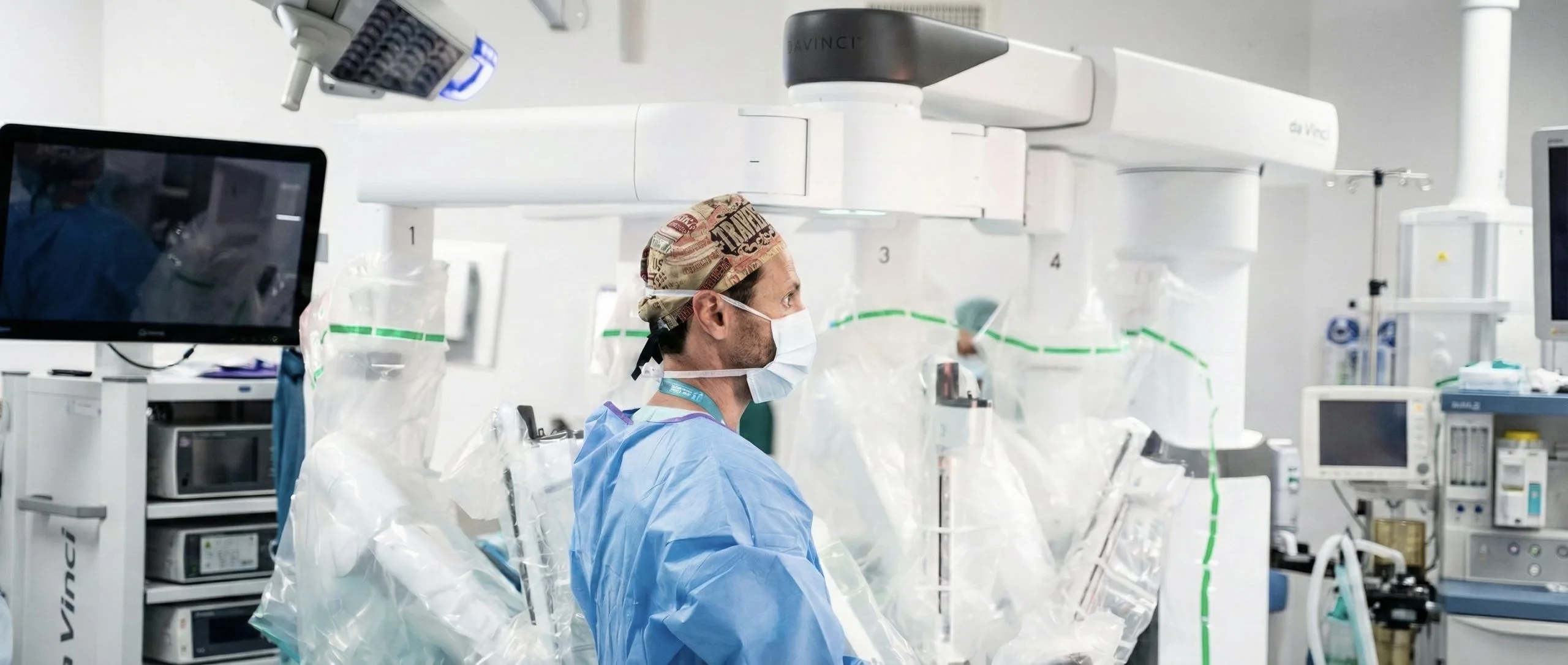
Robotic sacrocolpopexy (frequently using the Da Vinci® surgical system) is the most advanced way to perform this procedure via a minimally invasive route.
How it Works:
- It is performed under general anesthesia.
- Several small incisions (usually 4 to 5, about 0.5 to 1 cm each) are made in the abdomen.
- Through these incisions (“ports”), a 3D high-definition camera and miniaturized, highly articulated surgical instruments are inserted.
- The surgeon operates from an ergonomic console in the operating room, where they have a magnified three-dimensional view of the interior of the pelvis and control the robotic arms that manipulate the instruments with great precision.
Advantages over Other Approaches:
- Open Surgery: The robotic approach avoids the large abdominal incision, resulting in less pain, less blood loss, faster recovery, and a better aesthetic result.
- Conventional Laparoscopy: While also minimally invasive, conventional laparoscopy uses long, rigid instruments and offers a 2D view. Sacrocolpopexy involves delicate dissections in deep pelvic spaces and precise mesh suturing, which can be technically very challenging with conventional laparoscopy. Robotic surgery overcomes these limitations thanks to 3D vision, image magnification, tremor filtration, and, fundamentally, articulated instruments (EndoWrist®) that mimic and even exceed the movements of the human wrist.
4. Specific Advantages of Robotic Sacrocolpopexy
Robotic technology offers distinct benefits for performing sacrocolpopexy:
Magnified 3D High-Definition Vision: Allows exceptional visualization of complex pelvic anatomy, including the sacral promontory, vagina, bladder, rectum, ureters, and blood vessels, facilitating safe and precise dissection.
Greater Precision, Dexterity, and Range of Motion: Articulated robotic instruments allow the surgeon to perform fine and complex movements, essential for the meticulous dissection of the planes between the bladder and the vagina (vesico-vaginal space) and between the rectum and the vagina (recto-vaginal space), as well as for the precise suturing of the mesh to the vagina (or uterus) and to the sacral promontory ligament, even at difficult approach angles.
Meticulous and Safe Dissection: Facilitates the creation of the tunnels and spaces necessary for mesh placement, minimizing the risk of injury to adjacent structures.
Less Blood Loss During Surgery: And, consequently, a very low need for blood transfusions.
Less Post-operative Pain: Due to smaller incisions and less overall surgical trauma.
Shorter Hospital Stay: Generally, 1 to 3 days.
Faster Recovery and Earlier Return to Normal Activities and Work.
Better Aesthetic Results: Scars are small and discreet.
Excellent Long-term Anatomical and Functional Success Rates: The results of robotic sacrocolpopexy are comparable or even superior to open surgery, with the advantage of being minimally invasive.
Potentially Lower Risk of Mesh-Related Complications: Some studies suggest that the more precise fixation technique and reduced mesh manipulation may contribute to a lower risk of complications such as mesh erosion, although this point continues to be studied.
5. Who is a Candidate for Robotic Sacrocolpopexy?
Robotic sacrocolpopexy is an excellent option for many women with symptomatic apical prolapse, but the decision is always individualized. Ideal candidates generally include:
Women with symptomatic vaginal vault prolapse (the top of the vagina, after a previous hysterectomy).
Women with significant uterine prolapse (the uterus descends into or out of the vagina) who:
- Wish to preserve the uterus (in this case, a robotic sacrohysteropexy is performed, where the uterus is suspended).
- Or who will undergo a hysterectomy (removal of the uterus) during the same surgical procedure, followed by the suspension of the vaginal vault.
Frequently, women who also present prolapse of other pelvic compartments (such as cystocele – bladder prolapse – or rectocele – rectal prolapse), as the correction of the apical defect with sacrocolpopexy is fundamental to the overall success of pelvic floor repair.
Women who desire a durable repair of the prolapse and who want to maintain or resume vaginal sexual activity (sacrocolpopexy aims to restore the normal length and axis of the vagina, which is important for sexual function).
Women who are in adequate physical condition to tolerate general anesthesia and the specific surgical position (steep Trendelenburg position, with the head lower than the feet, to allow the intestines to move away from the pelvis and improve visualization).
It is important to discuss with your doctor all treatment options for prolapse, including alternatives to sacrocolpopexy (such as vaginal surgeries, for example, sacrospinous ligament suspension or colpocleisis) and non-surgical options (such as pessaries), to determine which is most appropriate for your specific case, considering your symptoms, lifestyle, expectations, and health status.
6. Preparation for Surgery
Preparation for robotic sacrocolpopexy includes:
Pre-anesthesia consultation.
Pre-operative exams: blood tests, urine analysis, ECG, etc.
Medication adjustment: suspending or adjusting medications such as anticoagulants or antiplatelet agents, according to medical guidance.
Fasting: as instructed.
Bowel Preparation: Frequently, a preparation to clean the bowel is recommended on the day before surgery to facilitate visualization and manipulation in the pelvis.
General information about hospitalization and what to expect.
7. The Surgical Procedure (Simplified Description for the Patient)
General anesthesia.
Surgical Positioning: Usually in lithotomy position with steep Trendelenburg.
Creation of ports and abdominal insufflation.
Robotic docking
Main Surgical Steps:
- Exposure of the Pelvis: The intestines are carefully moved away from the pelvis.
- Dissection of Vaginal Planes: The surgeon meticulously dissects the spaces between the bladder and the anterior wall of the vagina, and between the rectum and the posterior wall of the vagina. The sacral promontory is also exposed.
- Hysterectomy (if planned): If the uterus is present and it is decided to remove it (for example, a supracervical hysterectomy, where the cervix is preserved, or a total hysterectomy). If the uterus is preserved (sacrohysteropexy), the mesh will be fixed to the cervix or the uterine isthmus.
- Placement and Fixation of the Mesh: A synthetic mesh, usually in a “Y” shape or consisting of two pieces (anterior and posterior), is used. One portion of the mesh is sutured to the anterior wall of the vagina (and/or cervix/uterine isthmus) and another to the posterior wall. The “tail” of the mesh (or the upper ends of the two pieces) is then elevated and firmly fixed, without excessive tension, to the anterior longitudinal ligament over the sacral promontory, using sutures or special staples.
- Peritonization: The mesh is carefully covered with the peritoneum (the lining of the abdominal cavity) to isolate it from direct contact with the intestines and other pelvic organs, which helps prevent adhesions.
- Concomitant Procedures: If necessary, repairs of specific vaginal wall defects (e.g., correction of cystocele or rectocele with the patient’s own tissues – colporrhaphy) or surgery for stress urinary incontinence (e.g., placement of a urethral sling) may be performed.
Finalization: A catheter (bladder probe) is placed in the bladder. An abdominal drain may or may not be used. The small incisions are closed.
8. Post-operative and Recovery
Hospital Stay: Generally, the stay lasts from 1 to 3 days.
Pain Control: Post-operative pain is typically mild to moderate and well-controlled with analgesics.
Bladder Catheter: It is generally removed the day after surgery or before hospital discharge.
Abdominal Drain: If placed, it is removed when the amount of drained fluid is minimal.
Early Mobilization: You will be encouraged to get up and walk as soon as possible.
Diet: Feeding is restarted progressively.
Intestinal Function: It may take a few days to normalize. Mild laxatives may be prescribed to avoid strain.
Hospital Discharge: With detailed instructions on medication, wound care, and allowed physical activity.
Recovery at Home: Physical activity should be increased gradually. It is crucial to avoid lifting weights (more than 5 kg), making intense efforts, and having sexual intercourse for a period of about 6 to 8 weeks, or according to your doctor’s specific guidance, to allow for good healing and proper integration of the mesh into the tissues. It is important to prevent constipation. It is normal to feel some pelvic discomfort, fatigue, or a light vaginal discharge in the first weeks.
9. Expected Results and Potential Side Effects/Complications
Expected Results:
- High Anatomical Success Rates: Robotic sacrocolpopexy has very high success rates (generally over 90%) in correcting apical prolapse in the long term.
- Relief of Prolapse Symptoms: Such as the sensation of weight or pressure in the pelvis, the sensation of a “ball” in the vagina.
- Improvement of Bladder and Intestinal Function: If these were affected by the prolapse.
- Preservation or Improvement of Sexual Function: By restoring normal vaginal anatomy.
Potential Side Effects and Complications (most are rare):
- General Surgical Risks: Bleeding (rarely requiring transfusion), infection (wound, urinary, pelvic), deep vein thrombosis or pulmonary embolism (preventive measures are taken), injury to adjacent organs (bladder, ureter, rectum, large blood vessels – the risk is minimized with robotic precision and surgeon experience), port-site hernia.
- Specific Sacrocolpopexy Risks:
- Mesh-Related Complications (Though Infrequent with Current Techniques):
- Mesh Erosion:The mesh can, in rare cases, become exposed through the vaginal wall. The risk is low with modern macroporous, monofilament polypropylene meshes and careful placement techniques. If it occurs, it may require treatment with vaginal estrogen creams or, sometimes, a minor surgery to remove the exposed portion of the mesh.
- Chronic Pelvic Pain or Pain During Intercourse (Dyspareunia): It is rare, but can occur, sometimes related to the mesh, healing, or vaginal shortening if the technique is not optimized.
- Mesh Infection: This is a very rare but serious complication if it occurs, potentially requiring mesh removal.
- Constipation: May occur or worsen in some women after surgery.
- Low Back or Gluteal Pain (Sacralgia): Rare, may be related to the fixation of the mesh to the sacral promontory or to positioning during surgery.
- “De novo” Stress Urinary Incontinence (which did not exist before): Some women may develop urine leakage with effort after the prolapse is corrected, if the incontinence was “hidden” or “masked” by the prolapse itself. For this reason, your doctor will carefully evaluate your urinary function before surgery and discuss the eventual need for a concomitant anti-incontinence procedure.
- Mesh-Related Complications (Though Infrequent with Current Techniques):
- Prolapse Recurrence: Although success rates for apical sacrocolpopexy are very high, there may be, in the long term, a recurrence of prolapse in another vaginal compartment (e.g., anterior or posterior wall, if they were not specifically reinforced) or, very rarely, a failure of the apical suspension itself.
It is crucial that you openly discuss all these potential benefits and risks with your doctor, including those related to the use of surgical mesh, so that you can make a truly informed decision.
10. Medical Follow-up
After robotic sacrocolpopexy, you will have a regular follow-up plan with your doctor, which will include:
Review consultations to evaluate your recovery and healing.
Evaluation of the resolution of prolapse symptoms.
Pelvic exam to verify the anatomical success of the repair.
Long-term follow-up to monitor the durability of the repair and your general well-being.
11. My Experience with Robotic Sacrocolpopexy
“Robotic sacrocolpopexy is, in my clinical practice, the procedure I consider the ‘gold standard’ and my approach of choice for the surgical correction of apical prolapse (whether it is of the vaginal vault in previously hysterectomized women, or of the uterus in women who wish to preserve it – sacrohysteropexy) in patients seeking a long-lasting solution and who value the preservation of vaginal function. Robotic technology, with its superior three-dimensional vision, image magnification, and high-precision articulated instruments, allows me to perform this reconstructive surgery, which is complex and takes place in deep pelvic spaces, with remarkable safety and efficacy. Meticulous dissection of anatomical planes, precise placement and fixation of the polypropylene mesh (which I consider the most appropriate material for this suspension), and careful peritonization (covering the mesh with the abdominal lining) are crucial steps that are greatly facilitated by the robotic platform. My primary goal with each robotic sacrocolpopexy is to restore pelvic anatomy in the most physiological way possible, relieve symptoms durably, and substantially improve my patients’ quality of life, all with the recognized benefits of a minimally invasive approach.”
12. Final Message
Robotic sacrocolpopexy is an advanced, safe, and highly effective surgical treatment for prolapse of the top of the vagina or the uterus, offering excellent long-term success rates with the benefits of minimally invasive surgery. It allows for a robust anatomical correction, relieving symptoms and restoring quality of life. If you suffer from pelvic organ prolapse symptoms, such as the sensation of weight or pressure in the vagina, or a visible or palpable bulge, and this is affecting your daily life, robotic sacrocolpopexy may be the ideal solution for you. It is an important decision that should be carefully considered. I invite you to schedule a consultation for a full and personalized evaluation. We can discuss your specific case in detail, the potential benefits and risks of this procedure, and answer all your questions, so that you can make an informed and confident decision about your treatment.