Modern Treatment for Ureteropelvic Junction (UPJ) Obstruction
Brief Introduction:
If you or your child have been diagnosed with a Ureteropelvic Junction (UPJ) Obstruction – a blockage in the passage of urine from the kidney to the ureter – robotic pyeloplasty is one of the most advanced and effective surgical treatment options available today. This minimally invasive procedure aims to correct the obstruction, allow urine to flow normally, relieve symptoms, and, crucially, preserve renal function in the long term. This page was created to provide you with clear and detailed information about robotic pyeloplasty, helping you better understand this therapeutic option and make informed decisions together with your urologist.
1. What is the Ureteropelvic Junction (UPJ) and UPJ Obstruction? (A Brief Review)
Relevant Anatomy:
- Kidneys: Organs that filter the blood and produce urine.
- Renal Pelvis (or Bacinete): The central part of the kidney, funnel-shaped, where produced urine accumulates before passing to the ureter.
- Ureter: A thin muscular tube that actively carries urine from the renal pelvis of each kidney to the bladder.
- Ureteropelvic Junction (UPJ): The exact point where the renal pelvis narrows to continue as the ureter.
- UPJ Obstruction (UPJ Syndrome): Occurs when there is a narrowing or blockage at this junction, which prevents or hinders the normal passage of urine from the kidney to the ureter. As a result, urine accumulates “upstream” in the renal pelvis, causing its progressive dilation (a phenomenon called hydronephrosis). If not corrected, this obstruction can lead to pain, infections, stone formation, and, in the long term, damage to the function of the affected kidney.
2. What is Pyeloplasty?
Definition: Pyeloplasty is a reconstructive surgical procedure designed to correct the narrowing or blockage at the Ureteropelvic Junction (UPJ).
Standard Technique (Anderson-Hynes Dismembered Pyeloplasty): The most common technique with the best results involves the complete excision (removal) of the narrowed and abnormal segment of the UPJ. After removing this diseased area, the renal pelvis (which may be reduced in size if it is very dilated – reduction pyeloplasty) is carefully sutured (anastomosed) back to the ureter (which may also be “spatulated” – opened longitudinally to create a wider anastomosis opening), thus creating a new wide and functional junction. If the obstruction is caused or aggravated by an abnormal blood vessel that crosses and compresses the UPJ (polar or crossing vessel), this vessel is preserved, and the anastomosis is usually performed in front of it (ureteral transposition).
Main Objective: To restore normal and unobstructed drainage of urine from the kidney to the ureter, relieving hydronephrosis and symptoms (if present) and preventing progressive kidney damage.
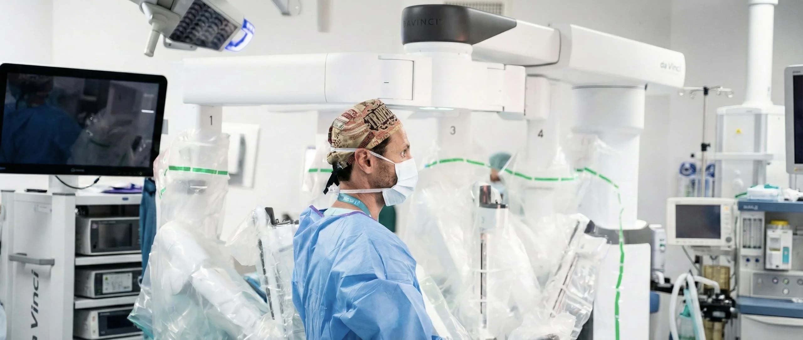
3. What is the Robotic Approach (Robot-Assisted Pyeloplasty)?
Pyeloplasty can be performed via traditional open surgery, conventional laparoscopy, or, more advanced and with multiple advantages, via the robotic route (frequently using the Da Vinci® surgical system).
How Robotic Surgery Works:
- It is a minimally invasive technique performed under general anesthesia.
- The surgeon makes several small incisions (usually 3 to 4, about 0.5 to 1 cm) in the patient’s abdomen.
- Through these incisions (“ports”), small tubes are inserted that allow the passage of a high-definition 3D camera and miniaturized, highly articulated surgical instruments.
- The surgeon operates from an ergonomic console in the operating room, where they have a magnified and detailed 3D view of the operative field.
- The surgeon controls robotic arms that hold the instruments.
- Hand movements on the console are translated in real-time into precise, filtered, and intuitive movements of the instruments inside the patient’s body.
- Open Surgery: Involves a larger incision in the flank (lateral back region), resulting in more post-operative pain, a larger scar, and a longer recovery. It was the standard approach in the past.
- Conventional Laparoscopy: Uses small incisions, but the surgeon operates with long, rigid instruments looking at a 2D monitor. The delicate and precise suturing required for pyeloplasty (the ureteropelvic anastomosis) can be technically very challenging with conventional laparoscopy due to restricted instrument movement and 2D vision.
- Robotic Surgery: Combines the benefits of a minimally invasive approach (small incisions) with cutting-edge technology that overcomes many of the limitations of conventional laparoscopy.Superior 3D vision, image magnification, tremor filtration, and, fundamentally, articulated instruments (EndoWrist®) greatly facilitate precise tissue dissection, excision of the narrowed segment, and, crucially, the performance of a delicate, precise, and watertight reconstruction (anastomosis) of the Ureteropelvic Junction.
Differences and Advantages over Other Approaches:
4. Advantages of Robotic Pyeloplasty
Robotic technology offers significant advantages for performing pyeloplasty, a delicate reconstructive procedure:
Magnified 3D High-Definition Vision: Allows exceptionally clear visualization of the UPJ anatomy, renal pelvis, ureter, and any abnormal blood vessels.
Greater Precision, Dexterity, and Range of Motion: Articulated robotic instruments allow the surgeon to perform fine and complex movements, essential for precise excision of the diseased segment and, more importantly, for creating a very precise and leak-proof suture (anastomosis) between the renal pelvis and the ureter.
Ease in Managing Crossing Blood Vessels: If a blood vessel is causing or contributing to the obstruction, robotic technology facilitates its careful dissection, preservation, and safe repositioning of the ureter (transposition) relative to the vessel.
Less Blood Loss During Surgery: Resulting in practically zero need for blood transfusions.
Less Post-operative Pain: Due to smaller incisions and less overall surgical trauma.
Shorter Hospital Stay: Generally, patients (especially children and young adults) can be discharged in 1 to 3 days.
Faster Recovery and Earlier Return to Normal Activities (including school or work).
Better Aesthetic Results: Significantly smaller and more discreet scars.
High Success Rates: Success rates in resolving the obstruction are comparable or even superior to traditional open surgery, generally above 90-95%.
5. Who is a Candidate for Robotic Pyeloplasty?
Robotic pyeloplasty is an excellent option for most patients (from infants to adults) diagnosed with significant Ureteropelvic Junction (UPJ) obstruction requiring surgical correction.
Indications for surgery generally include:
Presence of symptoms attributable to UPJ obstruction (such as flank or abdominal pain, recurrent urinary tract infections, nausea/vomiting associated with pain, or kidney stone formation).
Evidence of worsening hydronephrosis (increased kidney dilation) on serial imaging exams.
Demonstration of deterioration of renal function in the affected kidney (usually assessed by diuretic renal scintigraphy, showing relative function below 40% or a progressive decline in function).
In infants and young children, even if asymptomatic, surgery may be recommended if hydronephrosis is very pronounced or if there are signs of functional kidney compromise.
The decision to operate and the choice of technique are always individualized after a complete evaluation. The robotic approach is suitable for most cases of primary UPJ obstruction, including those associated with crossing vessels. It can also be used successfully in reoperation cases (e.g., after a previous failed pyeloplasty or endopyelotomy), although these situations can be more technically challenging.
6. Preparation for Surgery
Preparation for robotic pyeloplasty includes:
Pre-anesthesia Consultation: Evaluation by the anesthesiologist.
Pre-operative Exams: Blood tests, urine tests, ECG (if applicable), etc.
Medication Adjustment: Inform your doctor about regular medications, especially anticoagulants or antiplatelets, as they may need to be adjusted or temporarily suspended.
Fasting: Required for several hours before surgery.
General Information: Instructions regarding hospitalization, what to bring, etc.
7. The Surgical Procedure (Simplified Description for the Patient)
General Anesthesia.
Surgical Positioning: Usually, the patient is placed on their side (lateral decubitus) to allow access to the kidney and UPJ.
Creation of Ports: Several small incisions (ports) are made in the abdomen. The abdomen (or retroperitoneal space) is insufflated with CO2 to create a workspace.
Robotic Docking: Robotic arms with the 3D camera and instruments are introduced through the ports.
Main Surgical Steps:
- Exposure of the UPJ: The kidney, dilated renal pelvis, and transition zone (UPJ) are carefully dissected. Abnormal blood vessels (polar or crossing) are identified.
- Excision of the Obstructed Segment: The narrowed, non-functional segment of the UPJ is completely removed (excised). If the renal pelvis is very dilated, a reduction pyeloplasty may be performed.
- Ureteral Spatulation: The end of the ureter is opened longitudinally (spatulated) to create a wider opening for the new connection.
- Ureteropelvic Anastomosis (Reconstruction): This is the most critical stage. The renal pelvis is carefully sutured (anastomosed) to the spatulated end of the ureter using very fine sutures to create a new, wide, conical, and leak-proof junction. If there is a crossing vessel, the ureter is transposed so it is no longer compressed.
- Ureteral Stent Placement (Double J Catheter): Almost always, a thin, flexible internal catheter (Double J stent) is placed, extending from the renal pelvis, across the new anastomosis, down to the bladder. It serves as an internal mold for healing and ensures drainage.
Finalization: A small abdominal drain may be left (not always). Small incisions are closed.
8. Post-operative and Recovery
Hospital Stay: Generally short, ranging from 1 to 3 days.
Pain Control: Post-operative pain is typically mild to moderate and well-controlled with analgesics.
Urinary Catheter: A bladder catheter may be placed during surgery, usually removed the next day or shortly after.
Abdominal Drain: If placed, it is removed once drainage decreases (usually 1-2 days).
Early Mobilization: Encouraged to get up and walk as soon as possible.
Diet: Restarted progressively, usually on the day of or the day after surgery.
Recovery at Home: Activity should be increased gradually. Avoid intense efforts or contact sports for 4 to 6 weeks.
Double J Stent: Remains in place for several weeks (typically 4 to 6). During this time, it may cause discomfort or urinary symptoms (frequency, urgency, flank discomfort when urinating, or small amounts of blood in the urine). These symptoms are temporary and disappear after stent removal.
Stent Removal: Performed via a simple and quick endoscopic procedure (cystoscopy) under local anesthesia in an outpatient setting.
Imaging Follow-up: After surgery and stent removal, control imaging (ultrasound and later diuretic renal scintigraphy) will be performed to confirm success (resolution of hydronephrosis and improved drainage).
9. Expected Results and Potential Side Effects/Complications
Relief of Obstruction and Symptoms: The primary goal. Robotic pyeloplasty has success rates generally above 90-95%.
Preservation or Improvement of Renal Function: Aims to protect the kidney from future damage.
Potential Side Effects and Complications: Safe procedure, but like any surgery, has risks:
- General Risks: Bleeding (rarely requiring transfusion), infection (wound or urinary), deep vein thrombosis, injury to adjacent organs (bowel, spleen, pancreas – very rare with experienced robotic surgery), port-site hernia.
- Specific Pyeloplasty Risks:
- Urinary Fistula (Urine Leak): Leakage from the anastomosis. Usually resolves with keeping the drain/stent longer.
- Recurrent Obstruction or Pyeloplasty Failure: Although rare (<5-10%), narrowing can recur. May require new intervention (endoscopic or re-pyeloplasty).
- Double J Stent Complications: Discomfort, frequency, urgency, mild hematuria, infection, or rarely, encrustation/migration. Resolved upon removal.
- Kidney Stone Formation: Rare, but can occur if drainage is imperfect or if there is persistent infection.
10. Medical Follow-up After Robotic Pyeloplasty
The follow-up after surgery is important to ensure long-term success:
Regular consultations with the urologist.
Periodic Renal Ultrasounds: To monitor hydronephrosis resolution.
Control Diuretic Renal Scintigraphy: Usually performed 3-6 months after surgery to objectively confirm improved drainage and function.
Follow-up may be necessary for several years.
11. My Experience with Robotic Pyeloplasty
“Robotic pyeloplasty is, in my clinical practice, the treatment of choice and the standard of excellence for the surgical correction of ureteropelvic junction (UPJ) obstruction, in both pediatric and adult patients. This minimally invasive technique allows me to perform a precise and delicate anatomical reconstruction of the UPJ, with excellent and long-lasting functional results. The magnified high-definition 3D vision and superior dexterity of articulated robotic instruments are particularly advantageous for meticulous tissue dissection, precise excision of the narrowed segment, and, above all, the creation of a watertight and appropriately sized suture (anastomosis) between the renal pelvis and the ureter, even in the presence of abnormal blood vessels (polar vessels) that frequently complicate these cases. My primary goal with each robotic pyeloplasty is to provide my patients with effective and definitive relief of the obstruction, with maximum preservation of renal function, a fast and comfortable recovery, and minimal impact on their quality of life and that of their family.”
12. Final Message
Robotic pyeloplasty is a modern, safe, and highly effective surgical procedure for treating UPJ obstruction. It offers very high success rates combined with all the benefits of minimally invasive surgery, such as less pain, faster recovery, and better aesthetic results. If you or your child have been diagnosed with a UPJ obstruction requiring surgical correction, robotic pyeloplasty is, in the vast majority of cases, the best therapeutic option available today. I invite you to schedule a consultation so we can discuss your specific case in depth.